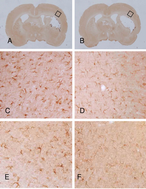
Figure 5.
PBBI-induced IL-1β immunoreactivity in vehicle (A) or NNZ-2566 (B) treated rats at 3 (C and D) or 7 days (E and F). IL-1β positive cells are visible in the region surrounding the lesion cavity at 3 days following PBBI (C) which are reduced following NNZ-2566 treatment (D). Panel E and F are representative photomicrographs of injured hemisphere at 7 days for PBBI (E) or PBBI + NNZ-2566 (F).

