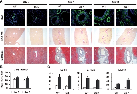
Figure 4.
A: Liver fibrosis was evaluated by α-SMA, Sirius red, and Masson’s trichrome staining 3, 7, and 14 days after BDL (Original magnification, ×200). B: Hydroxyproline content measured in the right median (#3) and the right lateral (#5) liver lobes 7 days after BDL. C: mRNA levels of transforming growth factor-β, α-SMA, and matrix metalloproteinase-3 were quantified by real-time RT PCR (sham operated mice are represented with open bars and BDL mice with closed bars).

