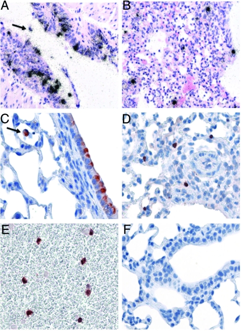
Figure 3.
Cell tropism in the respiratory tract and in peripheral blood. Visualization of virus RNA (A and B) and protein (C–E) in lungs of SC35M-infected mice 3 days p.i. by in situ hybridization and immunohistochemistry. Groups of five mice were infected with 106 PFU of recombinant virus. Numerous bronchial (A and C) epithelial cells as well as alveolar epithelial cells (B and D) were found to be positive early in infection. Also, mononuclear immune cells in the lung (A and C (arrows)) and in the blood (E) were found to be infected. Noninfected lungs were consistently negative in immunohistochemistry (F).

