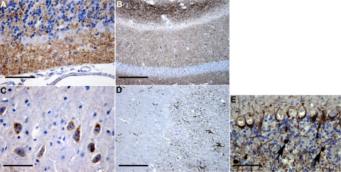
Figure 1.
Light microscopic pathology in Tg(PG14) mice. Sections from 361 to 394 day-old Tg(PG14) mice were labeled using antibodies against PrP (1A8) (A–C), glial fibrillary acidic protein (D), or phosphorylated neurofilament subunits (E). Diffuse PG14 PrP labeling was present in the molecular and granule cell layers of the cerebellum (A), in the hippocampus (B), and in perikaryonal cytoplasm of scattered neurons in the brainstem, including some in the locus coeruleus (C). PG14 PrP accumulation was accompanied by marked astrocytosis in the cerebellum (D) and by conspicuous axonal swellings containing phosphorylated neurofilaments in the cerebellar granule cell layer (arrows in E). Scale bars: 80–μm (A), 160–μm (B), 30–μm (C), 160–μm (D), and 80–μm (E).

