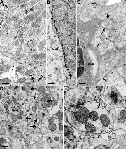
Figure 2.
Morphological changes in dendrites and glia associated with membrane labeling for PG14 PrP. Sections from Tg (PG14) mice at 361 to 394 days of age were examined by electron microscopy. A: Cerebellar granule cell layer showing individual puncta of PG14 PrP labeling, as well as short continuous segments of labeling (arrows) on the plasmalemmae of adjacent dendrites. B: Continuous labeling along a pyramidal neuron dendrite (den) and an adjacent unidentified dendrite from the stratum radiatum of the hippocampus. C: PG14 PrP labeling of dendrites is associated with an activated microglial cell (mic) and strands of microglial cytoplasm (arrows) invading between and around labeled processes. D: Unlabeled section from the cerebellar molecular layer showing thin strands of cytoplasm (arrows) from a microglial cell (mic) enveloping dendrites (den) and parallel fibers (asterisks). Parallel fiber axons show atrophy and excessive variation in cross-sectional profiles. E: Dendrite from an unlabeled section with an irregular, corrugated profile, part of which has a small out-folding of the plasmalemma (arrow). The dendrite is surrounded by excess extracellular space and fragments of tangentially sectioned processes. Scale bars: 0.3–μm (A), 0.15–μm (B), 0.3–μm (C), 0.45–μm (D), and 0.3–μm (E).

