Figure 3.
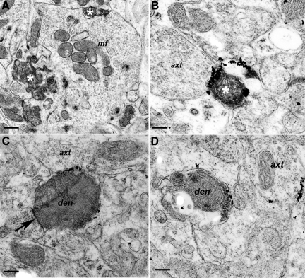
Dendrite degeneration associated with PG14 PrP labeling. Sections were from Tg (PG14) mice at 365 (A), 361 (B), 264 (C), or 361 (D) days of age. A: Two degenerate dendrites (asterisks), defined by their contacts with a mossy fiber terminal (mf), are shown in the granule cell layer of the cerebellum. B: A degenerate dendritic process (asterisk) in the cerebellar molecular layer showing marked PG14 PrP accumulation at its periphery. The process is in contact with an axon terminal (axt). C: Degenerate dendrite (den), as defined by the presence of an in-contact pre-synaptic density (arrow), with intense, focal, linear labeling on another pre-terminal axon (axt). D: An early-stage degenerating dendrite (den) with sparse membrane accumulation of PG14 PrP is shown. Nearby is an apparently healthy pre-terminal axon (axt), also with membrane PG14 PrP accumulation. Scale bars: 0.45–μm (A), 0.15–μm (B), 0.25–μm (C), and 0.20–μm (D).

