Figure 5.
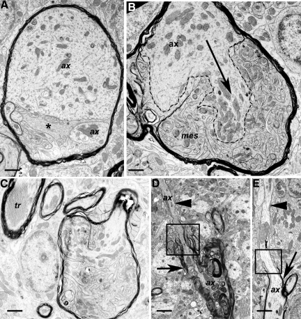
Pathology of myelinated axons and oligodendrocytes. Sections were from Tg (PG14) mice at 361 to 365 days of age. A: A swollen, myelinated axon divided into two parts (each labeled ax), the topmost of which contains large amounts of fine, fibrillar material, as well as endoplasmic reticulum and mitochondria. Mesaxonal proliferation is also present (asterisk). B: Axon (ax) showing marked proliferation or sprouting. One clearly defined sprout is indicated by the dashed lines and arrow. Prominent and highly complex proliferation of the inner mesaxon (mes) is also present, but the small size of the oligodendroglial and axonal sprouts makes it difficult to reliably distinguish between processes. C: A swollen myelinated axon with proliferation of the oligodendroglial, mesaxonal cytoplasm, and invasion of the axonal cytoplasm with partial compartmentalization by myelin. A myelinated process with a tubulo-reticular inclusion (tr) is adjacent. D: A degenerate myelinated process shows marked variation in myelin thickness. A thick myelin sheath is present on the lower part of the axon (arrow), with thin myelin on the upper part (arrowhead) above the paranode (square). The lower part of the axon is undergoing degeneration. ax; axon. E: An extended section of bare, unmyelinated axon (arrowhead) appears above the paranode (square) of a myelinated axon (arrow). ax, axon. Scale bars: 1.15–μm (A), 0.9–μm (B), 1.2–μm (C), 1.6–μm (D), and 1.6–μm (E).

