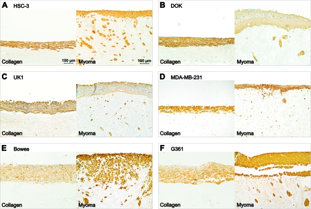
Figure 8.
Invasion of different cell types in organotypic cultures. Carcinoma or melanoma cells (7 × 10⁵) were cultured on top of type I collagen gel embedded with GF (7 × 10⁵) or on top of myoma tissue. Paraffin-embedded 14-day collagen and myoma organotypic sections were immunostained to visualize the cells. Tongue carcinoma cells HSC-3 (A), dysplastic oral keratinocytes DOK (B), and oral carcinoma cells UK1 (C) were stained for pancytokeratin AE1/AE3. D: Breast adenocarcinoma cells MDA-MB-231 were stained for E-cadherin. Melanoma cell lines Bowes (E) and G361 (F) were stained for S100. Scale bar: 100 μm.

