Figure 2.
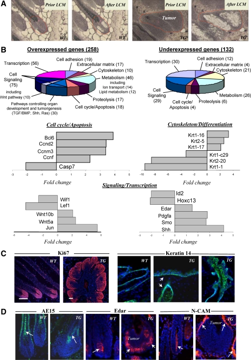
Hair follicle-derived tumors from TG mice show alterations in expression of cell cycle-associated genes and genes encoding pro-oncogenic molecules. Skin was harvested, and cryosections were processed for laser-capture microdissection (LCM), RNA isolation and amplification, microarray, and real-time PCR analyses, and for immunohistochemistry. A: HFs of wild-type (WT) mice and HF-derived tumors of TG mice before and after LCM. B: Diagrams showing the ontology of over- and underexpressed genes and real-time PCR analysis of genes differently expressed in the tumors versus normal hair matrix cells. C: Ki-67 expression in normal HFs and HF-derived tumors (left panel). Expression of Keratin 14 in the epidermis and HFs of wild-type and TG mice (right panel, arrows). D: Decrease in AE15 expression in the tumors of TG mice (left panel, arrow) versus normal anagen HFs of wild-type mice. Increase of Edar and N-CAM expression (central and right panels, respectively) in the tumor epithelium and mesenchyme of TG mice (arrows) versus wild-type HFs.

