Figure 4.
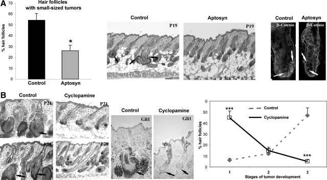
Treatment with the Wnt and Shh inhibitors differentially affects tumor formation in K14-Noggin mice. Back skin of Aptosyn- or cyclopamine-treated and nontreated mice was harvested at P19, P21, and P28, and processed for histoenzymatic visualization of alkaline phosphatase and histomorphometric analysis of HF-derived tumors. Statistical analyses were performed by using Student’s t-test. A: Significant decrease (*P < 0.01) in number of HFs with small tumors after Aptosyn treatment versus control. In control skin, small-sized HF tumors are shown by arrows (middle panel). In control HFs, nuclear β-catenin is seen in keratinocytes of the outer root sheath (left panel, arrows). In Aptosyn-treated mice, expression of nuclear β-catenin in the HF keratinocytes is strongly decreased (right panel, arrows). HFs are demarcated by dotted line. B: Significant decrease (***P < 0.001) in number of HFs bearing large and medium-sized tumors in TG mice treated by cyclopamine versus control (left and right panels, arrows). Decreased Gli1 expression in the HF keratinocytes after cyclopamine treatment (central panel, arrows).

