Figure 3.
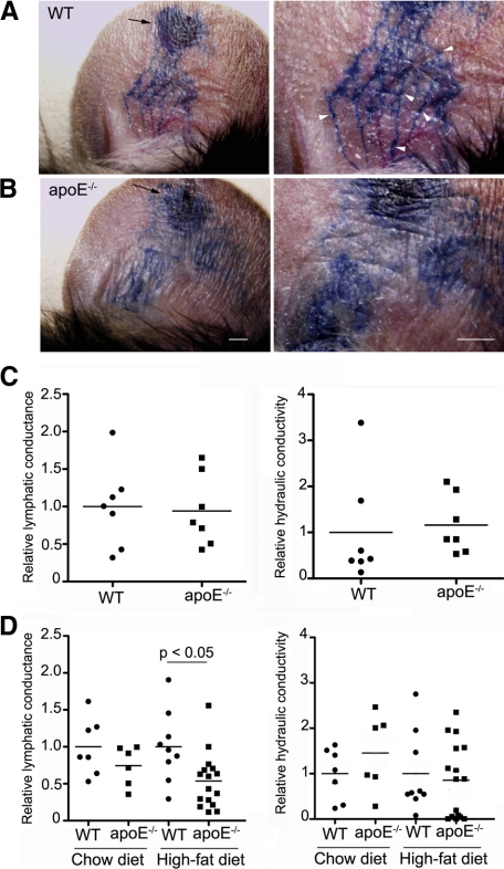
Impaired lymphatic vascular function in hypercholesterolemic mice. A, B: Evans blue dye was injected intradermally into the ear rim of 16- to 18-week-old wild-type (WT) and apoE−/− mice (arrows indicate the site of injection). After 1 minute, markedly dilated lymphatic vessels were visualized in apoE−/− mice ears, and Evans blue dye had extravasated from these dilated lymphatic vessels (B) compared with matched wild-type control mice, where fine lymphatic vessels were observed (arrowheads, A). C, D: Lymphatic uptake (conductance) and ease of fluid movement through the interstitial space (hydraulic conductivity) were analyzed by skin tail microlymphangiography in wild-type and apoE−/− mice at 4 to 5 weeks of age (C) or in older wild-type and apoE−/− mice maintained on a chow diet or fed a high-fat diet from 6 weeks of age (D). For each group, n ≥ 6.

