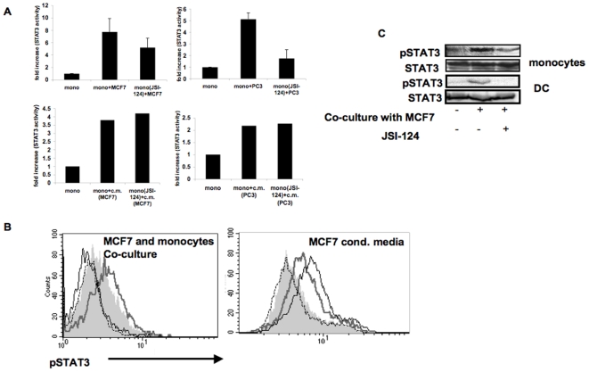
Figure 2. Contact-dependent but not conditioned media-induced STAT3 activity is sensitive to JSI-124 inhibition.
A. Monocytes were either left untreated or treated with JSI-124 (10 µM) for 1 hour at 37°C, and then washed. Treated and untreated cells were then cultured alone or were co-cultured with either MCF-7 or PC3 cells (upper panels) or with their conditioned media (lower panels). After 2 hours, nuclear extracts were prepared and tested for STAT3 activity using Non-Radioactive STAT3 Transcription Factor Assay. Comparable results were obtained in four separate experiments. B. Monocytes were either left untreated or treated with JSI-124 (10 µM) for 1 hour at 37°C, and then washed. JSI-124-treated and untreated cells were then cultured alone or co-cultured together with tumor cells (MCF-7) or with tumor cell's conditioned media. After 2 hours, cells were harvested and phosphorylated-STAT3 was detected by intracellular immunostaining and flow cytometric analysis. The two cell types were distinguished based on their forward and side scatter characteristics and phosphorylated STAT3 staining in monocyte population is shown. Dashed line: isotype control; Filled grey area: monocytes alone; Thick grey line: monocytes that were either co-cultured with MCF-7 (left panel) or were cultured in MCF-7 conditioned media (right panel); Black line: JSI-124-treated monocytes incubated with MCF-7 or their conditioned media, as above. Comparable results were obtained in three independent experiments. C. Monocytes or DCs were either left untreated or treated with JSI-124 (as indicated above), and then were co-cultured with MCF-7, for 2 hours. Untreated cells were obtained by mixing MCF-7 and either monocytes or DC without incubation. Cell extracts were subjected to SDS-PAGE and immunoblotting of anti-phosphorylated STAT3 demonstrate induction of STAT3 and that JSI-124 treatment blocks contact-dependent STAT3 phosphorylation. Anti-STAT3 immunoblotting reveals relative amounts of protein in each lane (lower panels).

