Figure 5.

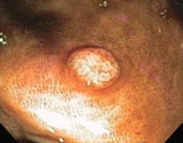
Easily visualized small sessile polypoid lesion in ascending colon with adjacent background pigmented mucosa typical of melanosis coli. Resected specimen confirmed absence of pigmented macrophages in the body of the resected adenoma.

Easily visualized small sessile polypoid lesion in ascending colon with adjacent background pigmented mucosa typical of melanosis coli. Resected specimen confirmed absence of pigmented macrophages in the body of the resected adenoma.