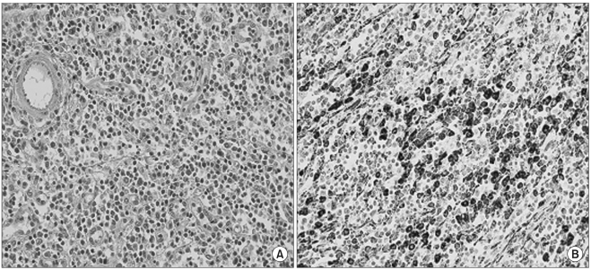
Fig. 2.
Left axillary lymph node biopsy. (A) Heavy infiltration of plasma cells and occasional eosinophils in the medullary cords and interfollicular areas (H & E, ×400). (B) Plasma cells were strongly reactive for Vs38, and some cells were large and multinucleated (Vs38 Immunohistochemical stain, ×400).

