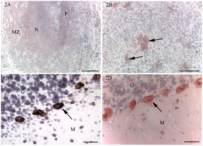
Figure 2. Immunohistochemistry for PrPC (A, B, D) and in situ hybridisation for PrP mRNA (C).
A. Spleen of control animal. A weak immunolabelling is present in a lymphoid nodule (N), the marginal zone (MZ) and PALS (P). B. Spleen of control animal. In the red pulp, immunolabelling for PrPC was mainly detected in single cells or small groups of cells (arrows). C, D. To correlate detection of PrPC and PrP mRNA, cerebellum of a normal one month old lamb was examined using immunohistochemistry and in situ hybridisation. C. In situ hybridisation. PrP transcripts were detected in Purkinje cells (arrow) and some scattered cells of the molecular (M) and granule cell (G) layers. D. PrPC immunohistochemistry. There is strong cytoplasmic immunolabelling of Purkinje cells (arrows) and scattered cells of the molecular (M) and granule cell (G) layers. Note the weak diffuse immunolabelling of the molecular layer that is not present with in situ hybridisation (Fig. 2C). Bars, 100 µm (A, B), 50 µm (C, D). Nuclei were stained with haematoxylin.

