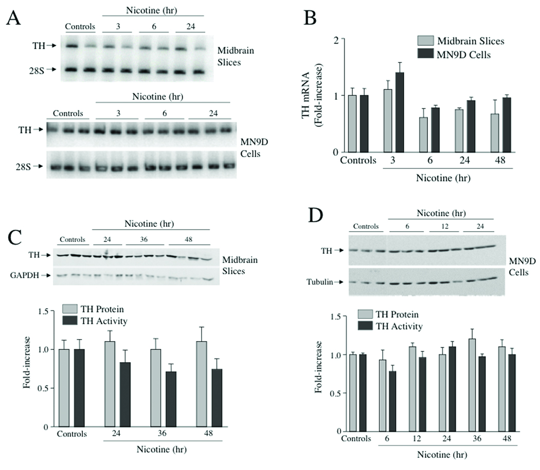
Figure 2. Nicotine did not induce TH mRNA, TH protein or TH activity in midbrain slice explant cultures or MN9D cells.
(A) Ventral midbrain slices from 7–10 day old mouse pups were cultured for 1 day in vitro and then treated with 100 uM nicotine for different periods of time up to 48 hr. MN9D cells were treated with 100 uM nicotine for similar durations of time. Total cellular RNA was extracted and semiquantitative RT-PCR was used to measure changes in TH RNA levels; 28S rRNA levels were also measured using this assay and these values were used for normalization purposes. The autoradiogram depicts representative assays of RNA isolated from either midbrain slice cultures or MN9D cells. (B) The bar graph depicts the means ± SE from 3 explant cultures or 3 dishes of MN9D cells. These results are from a single experiment, and similar results were obtained from at least two other experiments. (C) Midbrain slice explant cultures were treated with 100 uM nicotine for 24, 36 or 48 hr and then assayed for TH protein using western analysis and TH activity using saturating cofactor concentration (4 mM 6MPH4). The data represent means ± SE from 9–12 cultures. (D) MN9D cells were treated with 100 uM nicotine for the designated periods of time and TH protein and TH activity were assayed as in panel C. The data represent the means ± SE from 6–12 dishes.

