
Figure 1.
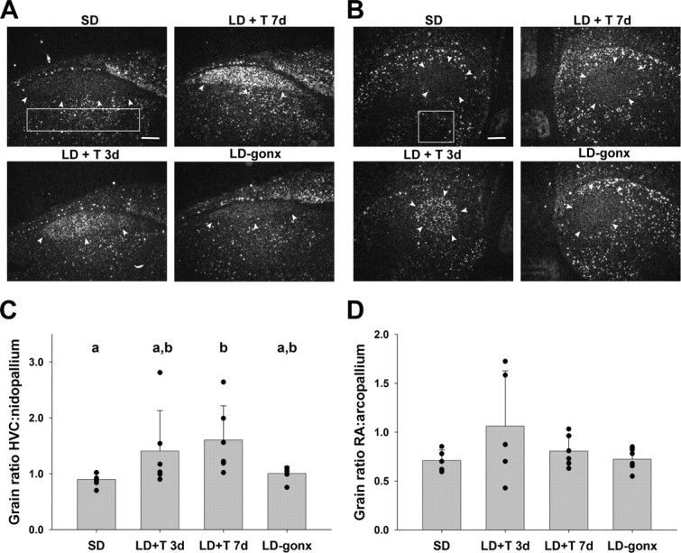
Dark-field photomicrographs of in situ hybridization showing BDNF mRNA expression in HVC (A) and RA (B) of birds in the four treatment groups. Arrows delineate the borders of the song nuclei. Boxes indicate area from which pallial measurements were taken. Scale bars, 300 μm. Quantitative analysis of BDNF mRNA expression in HVC (C) and RA (D). Data are expressed as a ratio of the grain density within the song nucleus versus the surrounding pallial region. Bars are group means ± SEM, and each point represents one bird. Letters above bars represent statistically different groups (Tukey's HSD p < 0.05).