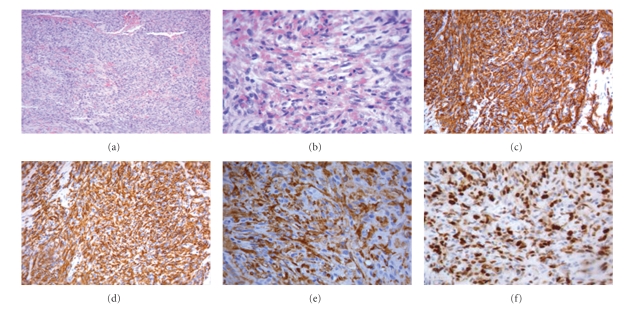
Figure 1.
(a) (colouring H&E 10×) Angiosarcoma moderately differentiated. (b) (colouring H&E 40×) Greater enlargement angiosarcoma moderately differentiated. (c) (20×) Immunohistochemical investigation tumor cells were positive for CD 31. (d) (20×) Immunohistochemical investigation tumor cells were positive for CD34. (e) (20×) Immunohistochemical investigation tumor cells were positive for FVIII rAg. (f) (40×) Positive for Mib-1 60%.

