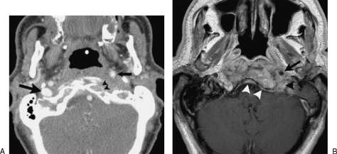
Figure 1.
Contrast-enhanced computed tomography (CT) of the skull base (A) shows erosion of the anterior cortex of the left occipital condyle (arrowheads) as well as anterior displacement of the left internal carotid artery (small arrow). The right internal jugular vein is normal (large arrow), whereas the left internal jugular vein is completely occluded. Gadolinium-enhanced magnetic resonance imaging (MRI) of the skull base (B) shows pathological contrast enhancement in the clivus extending into the soft tissues surrounding the left internal carotid artery (arrow) and jugular foramen (black arrowhead). There is thickening and enhancement of the clival dura (white arrowheads).

