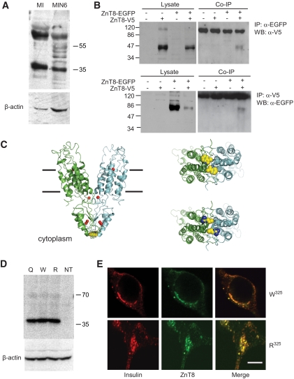
FIG. 6.
Effect of R325W polymorphism rs13266634 on the predicted molecular structure of ZnT8 and on subcellular localization and stability. A: Western immunoblotting of ZnT8 in mouse islets and clonal MIN6 β-cells. Total lysates were separated by 10% SDS-PAGE and immunoblotted with anti-rat/mouse ZnT8 polyclonal antibodies (Mellitech, France) or anti–β-actin polyclonal antibody (43 kDa; Sigma). B: Dimer formation. Human ZnT8-V5 and ZnT8-EGFP were transfected into HeLa cells. Forty-eight hours posttransfection, cells were lysed and subjected to immunoprecipitation with antibodies against EGFP (upper panel) or V5 (lower panel) tags. After washing the beads, bound proteins were eluted and subjected to immunoblotting, probing with anti-V5 (upper; predicted molecular mass for ZnT8-V5, 46.4 kDa) or anti-EGFP (lower; predicted molecular mass for ZnT8-EGFP, 68.2 kDa) antibodies. “NS” denotes a nonspecific band, likely derived from IgG lost from the sepharose beads. C: Modeling of ZnT8 structure based on YiiP. The human transporter was modeled using the structure of the Escherichia coli transporter YiiP (PDB accession 2QFI) as a template. Left, upper right: Provide views of the W325 variant from the plane of the membrane and the cytoplasmic side of the membrane, respectively; lower right, view of R325 variant from cytoplasmic side of the membrane. The likely locations of zinc ions are shown as red spheres in left, and residues at position 325 are shown in space-filling representation in all panels. D: Western immunoblotting analysis of overexpressed ZnT8 isoforms in MIN6 cells. E: Subcellular distribution of overexpressed ZnT8 isoforms in INS-1(832/13) cells; scale bar, 5 μm. (A high-quality digital representation of this figure is available in the online issue.)

