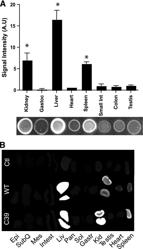
FIG. 3.
Adiponectin target organs. A: Dot-blot analysis of injected wild-type (WT) adiponectin in different organ lysates 4 h postinjection. Bar graph shows the signal intensity and below a representative dot blot. B: Representative direct infrared scanning of the full organs 4 h postinjection. Epi, epididymal white adipose tissue; Gast, gastrocnemius muscle; Kid, kidney; Liv, liver; Mes, mesenteric white adipose tissue; Pan, pancreas; Sol, soleus muscle; SubQ, subcutaneous white adipose tissue. n = 4.

