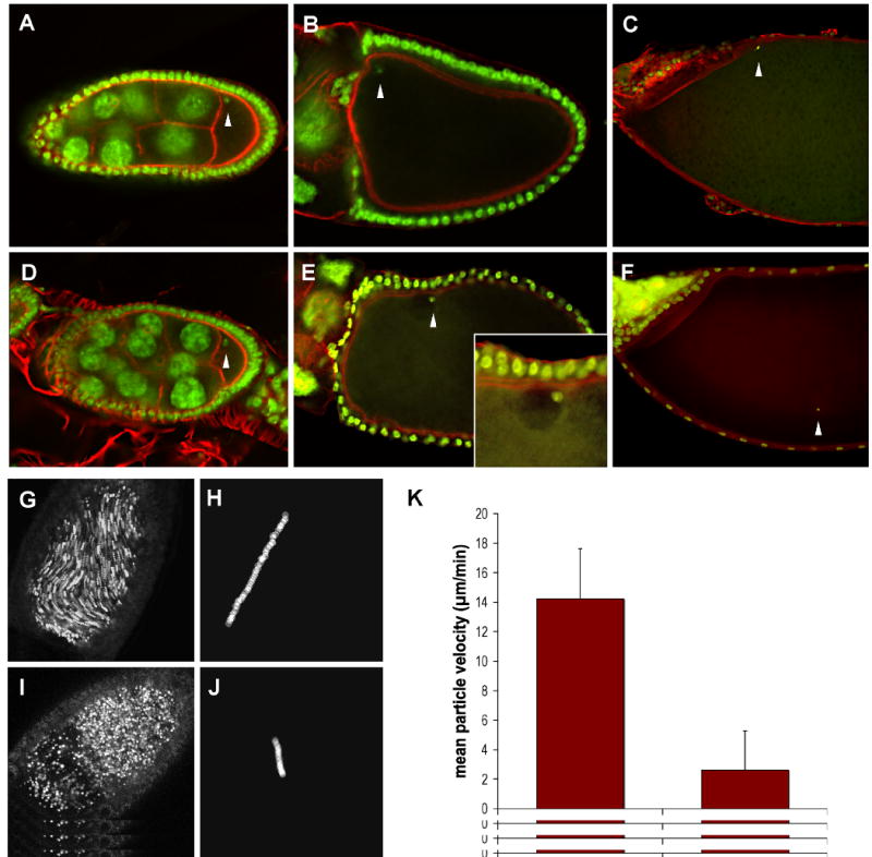
Fig. 1. Microtubule-dependent events are affected by msps mutations.

Oocytes from wild type (A-C) and mspsP/msps208 (D-F) females were fixed and stained with Sytox Green to reveal the position of the oocyte nucleus (arrowheads), and AlexaFluor-568 phalloidin to visualize actin filaments. The wild type nucleus is positioned in the anterior-dorsal corner of the oocyte, as evidenced by its position relative to the dorsal appendages. In the msps oocyte the nucleus is no longer anteriorly positioned after stage 10, although it appears to retain contact with the cell cortex (see inset in (E)).
Stage 10B oocytes from wild type (G) and mspsP/msps208 (I) females were dissected into halogen oil 27 and imaged using the fluorescein channel of a Nikon confocal microscope. Five consecutive images were collected at ten-second intervals and merged. Particles whose position has changed over the time course of the experiment appear as streamers. Directional movement is evident in the wild type oocyte but not the msps oocyte, which exhibits the stratified cytoplasm commonly observed in these mutants. (H,J) The movement of a single particle was tracked over 500sec/50 frames. Individual particle tracings were used to calculate average particle velocities for wild type and mspsP/msps208 stage 10B oocytes (see text). (K) Time lapse stacks were used to calculate average particle velocities for wt and mspsP/msps208 stage 10b oocytes (n=20 for each genotype) (bottom). Bars indicate standard deviation.
