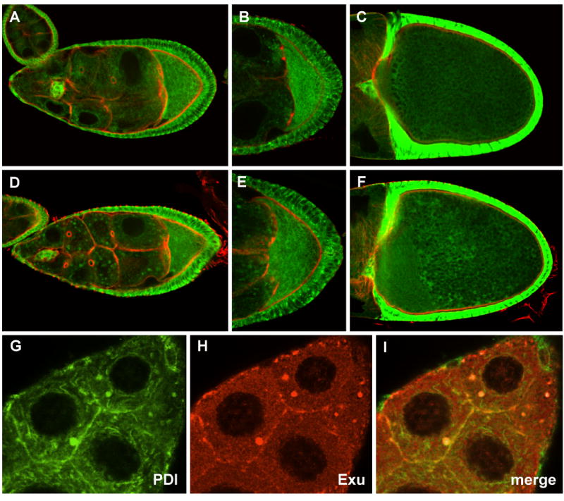
Fig. 4. Expression of PDI-GFP in wild type and msps egg chambers.

PDI-GFP distribution in wild type (A-C), or msps (D-F) egg chambers (counterstained with phalloidin (red)). During stage 9 (A, B) PDI is distributed in a mesh-like array in the nurse cells, and is present at higher concentrations in the oocyte. Msps mutations (D,E) result in minor perturbations in PDI-GFP distribution. At stage 10 PDI is uniformly distributed in wild type oocytes (C), but its distribution becomes somewhat irregular in msps mutants (F). (G-I) The distribution of PDI-GFP (G) and Exu (H) in the nurse cell cytoplasm of a stage 9 msps egg chamber were compared. Only partial overlap in signal is observed (I).
