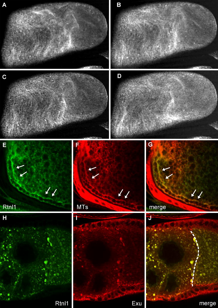
Fig. 6. Co-localization of Rtnl1 with microtubules and Exu.

Fibers in the cytoplasm of a wild type stage 11 oocyte are decorated with Rtnl1-GFP (A-D) and move with a ripple-like motion in these live images, taken 40 seconds apart. Wild type stage 10B oocytes expressing Rtnl1-GFP (E) were stained for tubulin (F). Rtnl1 co-localizes with microtubules in streaming oocytes (G). The arrows indicate parallel subcortical microtubules typical of this stage. msps egg chambers expressing Rtnl1-GFP (H) were stained with antibodies to Exu (I). Exu colocalizes with Rtnl1 in msps mutants (J). The dashed line in (J) indicates the nurse cell-oocyte border.
