
Figure 5.
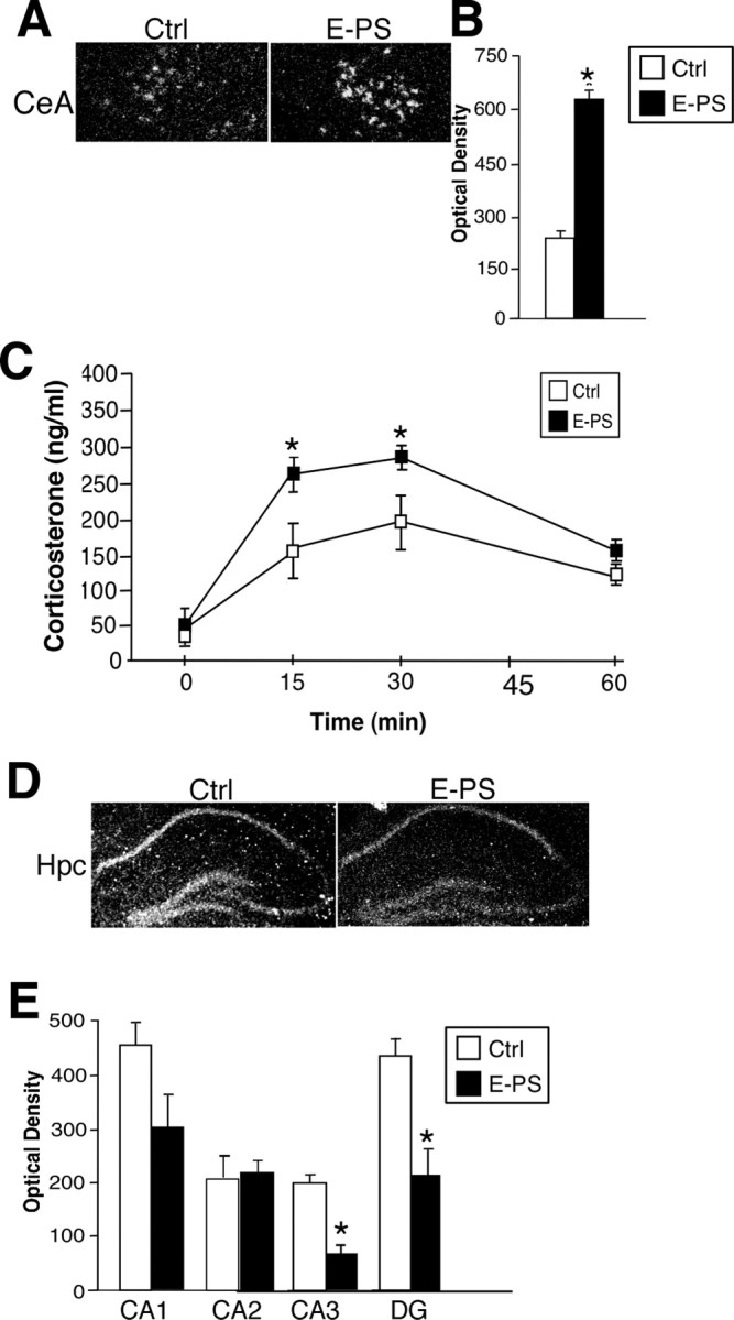
E-PS males displayed stress pathway dysregulation including increased CeA CRF, decreased hippocampal GR, and an increased HPA axis stress response. A, B, E-PS significantly increased CRF expression in the central nucleus of the amygdala compared with controls (*p < 0.05; n = 4). C, Corticosterone levels after a 15 min restraint stress were significantly higher in E-PS males compared with controls (*p < 0.05; n = 6). D, E, GR expression was significantly lower in E-PS males compared with controls in the CA3 and DG of the dorsal hippocampus (*p < 0.05). The apparent decrease in CA1 GR expression was not significant (p = 0.07; n = 5). No effect of E-PS was seen in CA2. Data are mean ± SEM. Hpc, Hippocampus.