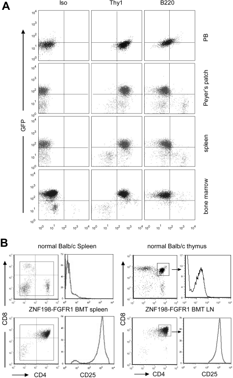
Figure 2.
Immunophenotype of hematopoietic cells of the diseased mice. (A) Flow cytometric analysis of different tissue samples, showing high levels of GFP+ Thy1+ cells in all tissues analyzed. (B) The majority of splenocytes and lymphocytes in leukemic mice are CD4 and CD8 DP, with high expression of CD25, compared with CD4+/CD8+ cells isolated from normal thymus.

