
Figure 4.
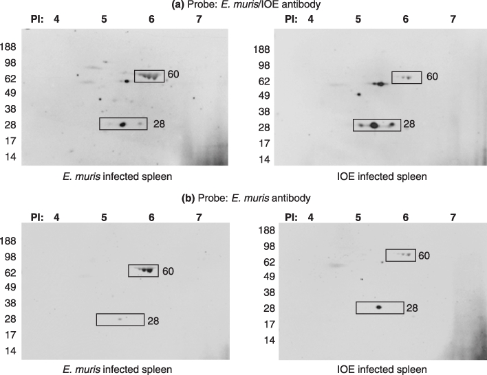
Western blot of two dimensional gels (E. muris infected spleen and IOE infected spleen) probed with polyclonal antibodies against (a) E. muris/IOE and (b) E. muris. Fifty micrograms of protein was used to run the first dimensional gel. The proteins of interest are marked in the image (rectangle).