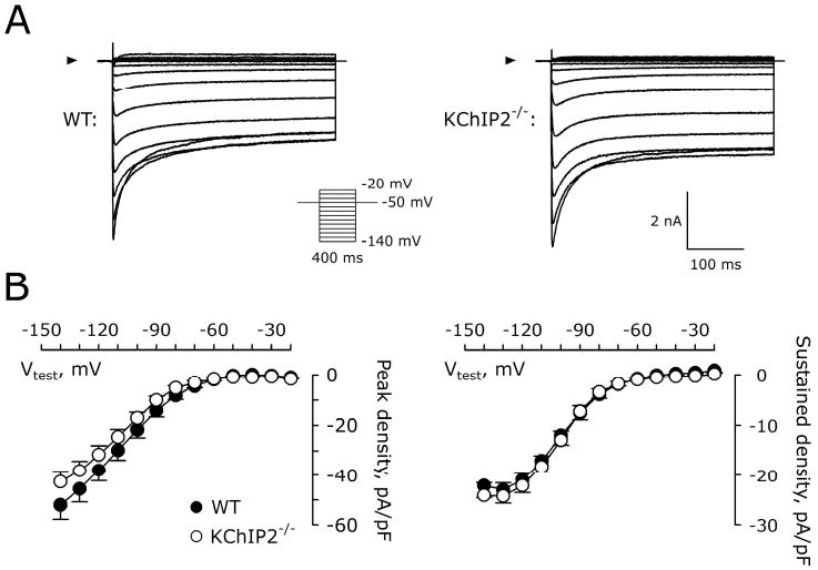
Figure 3.

Similar inward rectifying K+ currents in WT and KChIP2-/- myocytes. A, Representative current traces recorded from a WT and KChIP2-/- myocyte. Arrowheads indicate 0 pA. Inset, voltage protocol. B, Peak inward IK1 (left) and steady state (right) IK1 in WT (n=7) and KChIP2-/- (n=10) myocytes.
