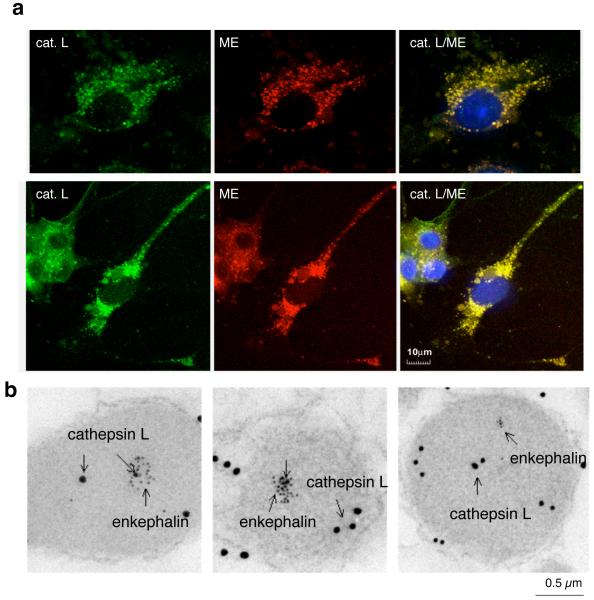
Figure 6. Localization of cathepsin L to neuropeptide-containing secretory vesicles.
(a) Colocalization of cathepsin L with enkephalin in chromaffin cells demonstrated by confocal immunofluorescence microscopy. Cathepsin L and (Met)enkephalin (green and red fluorescence, respectively) in chromaffin cells were visualized by immunofluorescence confocal microscopy. Excellent colocalization of cathepsin L and (Met)enkephalin was demonstrated by the merged images with colocalization indicated by yellow fluorescence. In chromaffin cells, the majority of cathepsin L is colocalized with (Met)enkephalin within secretory vesicles.
(b) Immunoelectron microscopy demonstrates colocalization of cathepsin L with the (Met)enkephalin neuropeptide in secretory vesicles. Cathepsin L localization was indicated by labeling with 15 nm colloidal gold-conjugated anti-rabbit, and (Met)enkephalin (ME) was detected as 6 nm gold particles conjugated to anti-mouse. The presence of both 15- and 6-nm cold particles within these vesicles demonstrated the colocalization of cathepsin L with the enkephalin neuropeptide in secretory vesicles.

