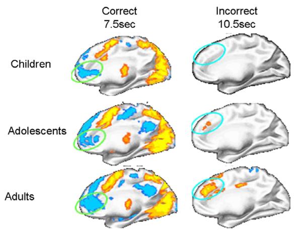
Fig. 5.
Activation maps displayed on the partially inflated medial cortical surface of the right hemisphere for inhibitory errors in the AS task for children, adolescents, and adults. Results indicate similarities across age groups during the initial stage of error processing in the medFG/rACC. However, only adults show recruitment of dACC in later stages of error processing. Blue indicated deactivation. Red/Yellow indicated activation (adapted from Velanova et al. (2008). Cerebral Cortex, February 14 [Epub ahead of print]). (For interpretation of the references to color in this figure legend, the reader is referred to the web version of this paper.)

