
Figure 4. Growth Suppression of Embryonal Carcinoma Cells by miR-200c.
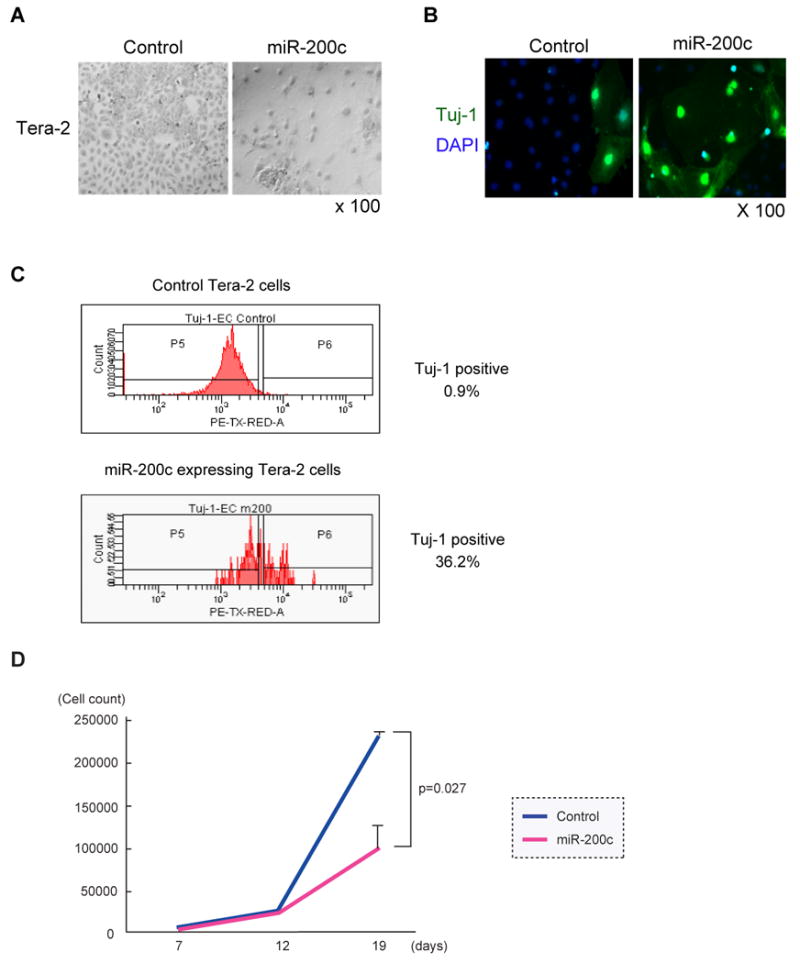
(A) Images of miRNA-expressing embryonal carcinoma cells. Tera-2 cells infected with the miRNA expressing lentivirus were collected by flow cytometry 4 days after infection. Tera-2 cells were cultured for 19 days and stained with Giemsa Wright staining solution. (B) MiR-200c enhanced differentiation of embryonal carcinoma cells. Tera-2 cells as described in (A) were stained with primary antibody against the early post-mitotic neuron marker, Tuj1 followed by Alexa-488 labeled secondary antibody. Cells were counterstained with DAPI. (C) Flow cytometry analysis of Tuj-1 expression. Tera-2 cells infected by miR-200c expressing lentivirus or control lentivirus were cultured for 6 days. Tera-2 cells were permeabilized and stained by anti-Tuj-1 antibody. Tuj-1 expression of GFP expressing Tera-2 cells was analyzed by flow cytometry. (D) MiR-200c inhibited the growth of embryonal carcinoma cells in vitro. 3000 miR-200c expressing or control Tera-2 cells were collected as described in (A) and cultured in a 96-well plate. Total cell numbers were counted on days 7, 12 and 19. The result is the average and S.D. from three independent wells.