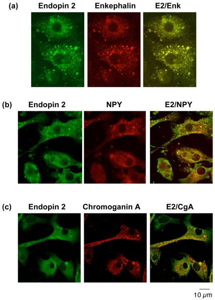
Figure 4. Endopin 2 colocalization with neuropeptides in secretory vesicles of chromaffin cells demonstrated by immunofluorescence confocal microscopy.
a. Endopin 2 and enkephalin. Immunofluorescence colocalization of endopin 2 (E2) with the enkephalin neuropeptide in chromaffin cells (bovine) was assessed by rabbit anti-endopin 2 detected with anti-rabbit IgG-Alexa Fluor 488 (green fluorescence), and detection of (Met)enkephalin (Enk) with mouse anti-Enk detected with anti-mouse IgG Alexa 594 (red fluorescence). The colocalization of endopin 2 and (Met)enkephalin is illustrated by overlay of the images, illustrated by yellow fluorescence.
b. Endopin 2 and NPY. Immunofluorescence colocalization of endopin 2 (E2) with the NPY neuropeptide in chromaffin cells (bovine) was assessed by rabbit anti-endopin 2 detected with anti-rabbit IgG Alexa Fluor 488 (green fluorescence), and detection of NPY with sheep anti-NPY detected with anti-sheep IgG Alexa 594 (red fluorescence). The colocalization of endopin 2 and NPY is illustrated by overlay of the images, illustrated by yellow fluorescence.
c. Endopin 2 and chromogranin A. Immunofluorescence colocalization of endopin 2 (E2) with the secretory vesicle marker chromogranin A (CgA) in chromaffin cells was assessed by rabbit anti-endopin 2 detected with anti-rabbit IgG Alexa Fluor 488 (green fluorescence), and detection of CgA with mouse anti-CgA detected with anti-mouse IgG Alexa 594 (red fluorescence). The colocalization of endopin 2 and CgA is illustrated by the overlay of the images, illustrated by yellow fluorescence.

