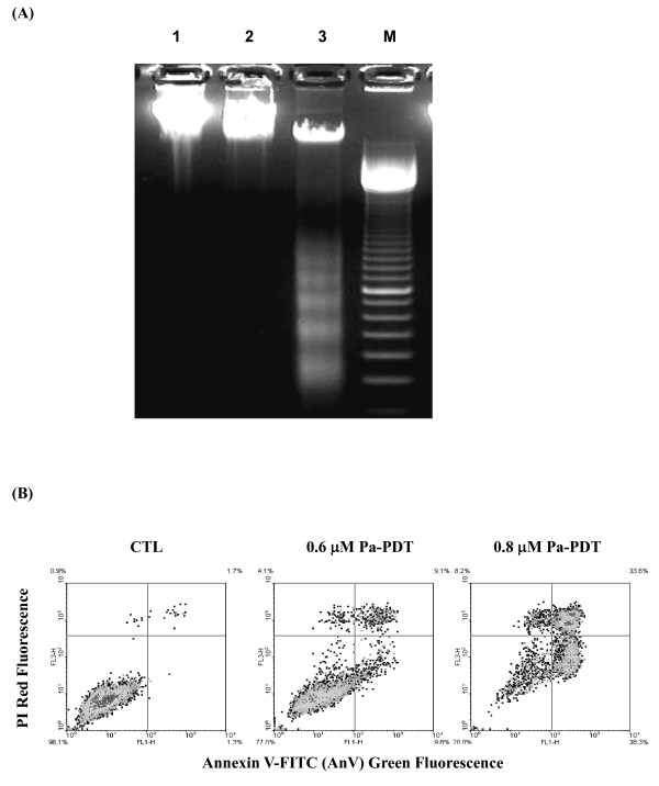
Figure 2.
Pa-PDT induces apoptosis in R-HepG2 cells. (A) DNA fragmentation was revealed by agarose gel electrophoresis. The cells (2 × 10⁶/plate) were collected at 24 h after the treatment, Lane 1: solvent control (0.04% ethanol with light illumination); lane 2: cells treated with Pa (0.6 μM) without light illumination; lane 3: cells treated with Pa (0.6 μM) with 20 min light illumination (84 J/cm2); and lane M: DNA markers (100 base pair). (B) For the annexin V/PI assay, the Pa-PDT treated cells were collected at 2 h and then stained with annexin V-FITC and PI (15 min) for apoptosis study. Results shown are representative of 3 independent experiments.

