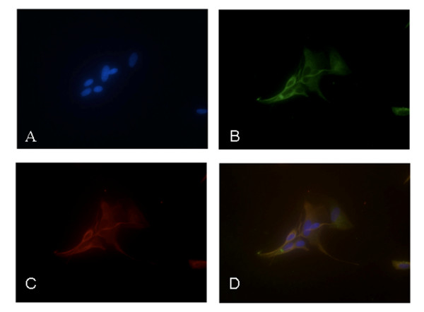
Figure 2.
Immunostaining of primary cultures of trigeminal satellite cells. Cells were stained with the nuclear dye DAPI (A). Immunocytofluorescence was performed with antibodies against the glial fibrillary acidic protein (GFAP) (B) and glutamine synthase (GS) (C). The same field is shown in all three panels, and a merged representation of the triple-staining is presented in panel D. (40× – 50 μm scale bar)

