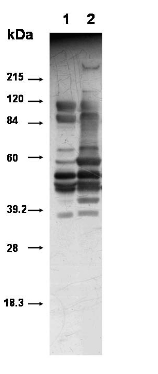
Figure 4.
Western blot analysis of immunogenic proteins of whole cell lysate of C. perfringens grown on TPYG (lane 1) and CMM (lane 2). Protein was separated on 12% SDS-PAGE and transferred onto PVDF membrane. Mouse anti- C. perfringens serum (obtained from animals that survived experimental gas gangrene infection) was used to probe the blot and bound antibodies were detected by Goat anti-mouse IgG HRP conjugate by chemiluminescence using and ECL western blot kit (Sigma).

