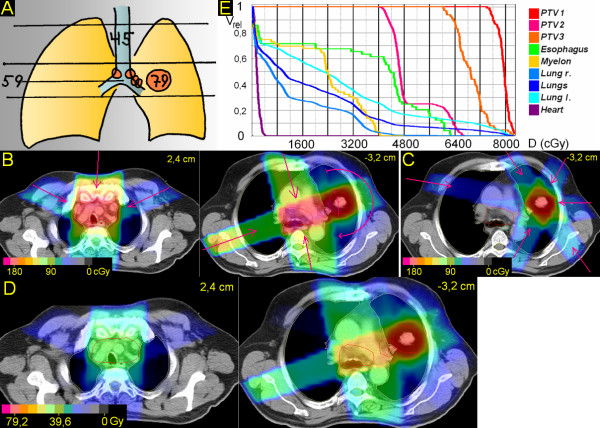
Figure 5.
Peripheral tumor located lateral, but close to hilus/mediastinum. 67 years; adenocarcinoma peripheral upper lobe, 3,5 cm ∅; enlarged hilar nodes, mediastinoscopically proven bilateral mediastinal nodes. A. Scheme; position of junction plane and upper and lower borders, doses (Gy). B. Treatment plan single fraction. C. Treatment plan single fraction of boost to primary tumor. D. Overall treatment plan. E. DVHs.

