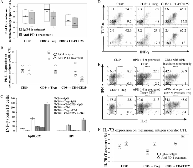
Fig. 4.
PD-1 blockade augmented the function of melanoma antigen-specific CTLs and reversed the impairment of effector function by Treg. (A) PD-1 blockade reduced PD-1 expression on melanoma antigen-specific CTL induced by Treg; + represented the median and − represented the mean PD-1 expression on melanoma antigen-specific CTL. Data were summarized from five separate experiments. (B) PD-1 blockade decreased PD-L1 expression on melanoma antigen-specific CTL up-regulated by Treg; + was the median and * was the mean of PD-L1 expression on melanoma antigen-specific CTL. The data shown as scatter dot plots from four individual experiments. (C) Anti PD-1 antibody reversed the impairment of INF-γ secretion by CTL induced by Treg. INF-γ secretion was presented as mean ± SD. This figure showed one representative experiment from six. (D) The impact of PD-1 blocking antibody on intracellular INF-γ and TNF-α secretion by melanoma antigen-specific CTL. The upper row represents cultures treated with control antibody and the lower row was the cultures treated with PD-1-blocking antibody for 10 days. (E) PD-1 blockade increased intracellular INF-γ and IL-2 co-expression in melanoma antigen-specific CTLs under different culture conditions. Dot plot is gated on tetramer gp100-2M+CD8+ T cells. (F) PD-1-blocking antibody treatment up-regulated the IL-7R expression on melanoma antigen-specific CTL; + was the median and * was the mean of the IL-7R expression on melanoma antigen-specific CTL. Data were shown as scatter plots from three individuals experiments.

