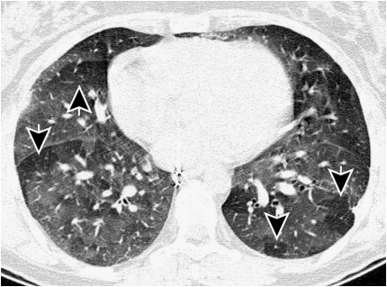
Figure 3.
This figure shows a thin slice (1 mm slice thickness) CT scan acquired after expiration. Most of the lung has increased density (less dark), but parts of the lung are exhibiting signs of gas trapping (arrows) and appear darker on the image. It is thought that these regions of gas trapping are caused by small airway narrowing.

