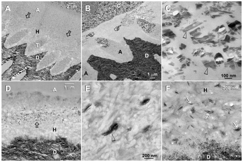
Fig. 1.

TEM images taken from unstained, undemineralized sections of resin-bonded dentin that were immersed in the first experimental Portland cement/SBF remineralization medium containing PAA as the only biomimetic analog. Abbreviations: C: composite; A: adhesive; H: hybrid layer; T: dentinal tubule; D: mineralized intertubular dentin. A. A representative 4-month old specimen that was bonded with XP Bond using the moist bonding technique. There was no remineralization when PVPA was absent. Presumably, the hybrid layer was better infiltrated than the dry bonding group [see Fig. 1B], and there were no spaces to accommodate the deposition of large crystals. Open arrows: top of the hybrid layer. B. A 4-month old specimen that was bonded with XP Bond using a dry bonding technique. A cluster of large crystals (pointers) could be seen within the hybrid layer. C. A high magnification view of Fig. 1B. Many of the crystals that were formed in the absence of PVPA were longer than 100 nm along their C-axes (arrowheads). They were too large to fit inside collagen fibrils (i.e. intrafibrillar remineralization) and were probably formed around the interfibrillar spaces. D. Adper Prompt L-Pop-bonded dentin that was retrieved after 4 months of immersion in the PAA-containing remineralization medium. A zone of electron-dense crystals appeared in the middle of the hybrid layer. E. A high magnification view of electron-dense amorphous structures (open arrowhead) that appeared within the hybrid layer of Adper Prompt L-Pop-bonded dentin after immersion in the same remineralization medium for 2 months (low magnification not shown). F. A high magnification of Figure 1D. At 4 months, the electron-dense amorphous structures depicted in Fig. 1E had been converted to crystalline plates (arrows) that were 100–150 nm in length along their C-axes. These remineralized crystals were considerably larger than the natural apatite crystallites in the mineralized dentin base.
