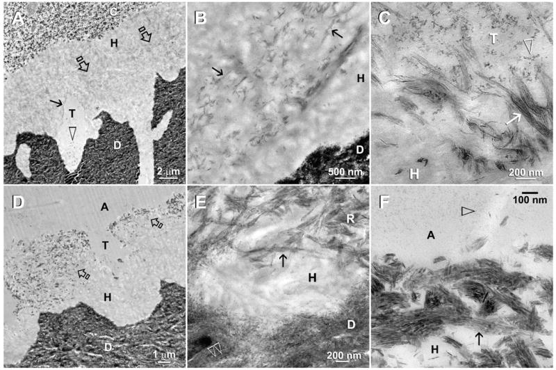
Fig. 2.

TEM of moist-bonded XP bond specimens that had been immersed in the second experimental PAA- and PVPA-containing remineralization medium. The specimens were examined after 2 months (A–C) and 4 months (D–F) of biomimetic remineralization. Abbreviations: C: composite; A: adhesive; H: hybrid layer; T: dentinal tubule; D: mineralized intertubular dentin. A. In this two-month old specimen, remineralization could be vaguely discerned from the middle of the hybrid layer (open arrows) and around the periphery of the dentinal tubule (arrow). Adhesive nanofillers (open arrowhead) were found within a dentinal tubule. B. A moderately high magnification view of Fig. 2A showing regional intrafibrillar remineralization (arrows) of the resin-infiltrated collagen matrix. Presumably, the adjacent interfibrillar spaces were well-infiltrated by resin and lacked spaces for mineral deposition. C. High magnification of Fig. 2A showing the close proximity between adhesive nanofillers (arrowhead) and the partially-remineralized collagen fibrils (arrow) along the periphery of a dentinal tubule. These fine intrafibrillar minerals recapitulated the rope-like subfibrillar architecture of the collagen fibrils. As these fibrils were probably swollen and partially-unraveled, no collagen banding was observed. D. At 4-months, remineralization within the hybrid layer (open arrows) could be identified even at a low magnification. E. A moderately high magnification view of Fig. 2D showing remineralized collagen fibrils (R) close to the base of the hybrid layer. Although the hierarchy of intrafibrillar mineralization had been recapitulated (arrow) in these fibrils, the crystallite arrangement lacked the banded characteristic of mineralized fibrils (open arrowheads) present in the underlying dentin. F. Remineralized collagen fibrils along the hybrid layer surface. Intrafibrillar mineral nanoplatelets (arrow) were 50–70 nm long and their arrangement reflected the pleaded, rope-like arrangement of the collagen microfibrils. Open arrow: adhesive nanofillers.
