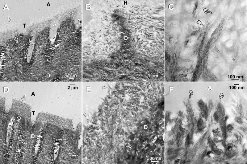
Fig. 4.

TEM of Adper Prompt L-Pop bonded specimens that that had been immersed in the second experimental PAA- and PVPA-containing remineralization medium. The specimens were examined after 2 months (A–C) and 4 months (D–F) of biomimetic remineralization. Abbreviations: C: composite; A: adhesive; H: hybrid layer; T: dentinal tubule; D: mineralized intertubular dentin. A. A low magnification view of a two-month old specimen showing a 4–6 μm thick hybrid layer (between open arrows) with remineralization of the bottom part the hybrid layer (asterisk). Other specimens revealed remineralization initiating from the top and the hybrid layer (not shown). B. A moderately high magnification view of a less heavily remineralized specimen from the same time period. Remineralization was seen exclusively as intrafibrillar remineralization within some collagen fibrils (open arrowheads). C. A high magnification view of the collagen matrix at the base of the hybrid layer showing part of a collagen fibril that had undergone intrafibrillar remineralization (open arrowhead) and another non-mineralized region within the same fibril (pointer). The remineralized part of these fibrils consisted of orderly arranged mineral nanoplatelets, revealing the rope-like subfibrillar architecture within these collagen fibrils. D. Low magnification of a four-month old specimen showing partial remineralization of the entire 4–6 μm thick hybrid layer (between open arrows). E. A moderately high magnification view of Fig. 4D showing remineralization within the hybrid layer (between open arrows) that is principally intrafibrillar in nature. Highly remineralized shag carpet-like fibrils (pointers) could be indentified along the dentin surface. Presumably, the adhesive resin-infiltrated interfibrillar spaces did not remineralize and appeared electron-lucent. F. A high magnification view of the heavily remineralized collagen fibrils along the hybrid layer surface. The extent of surface unraveling in these collagen fibrils (pointers) could be readily appreciated by comparing the width of these fibrils from a subsurface location (between open arrowheads).
