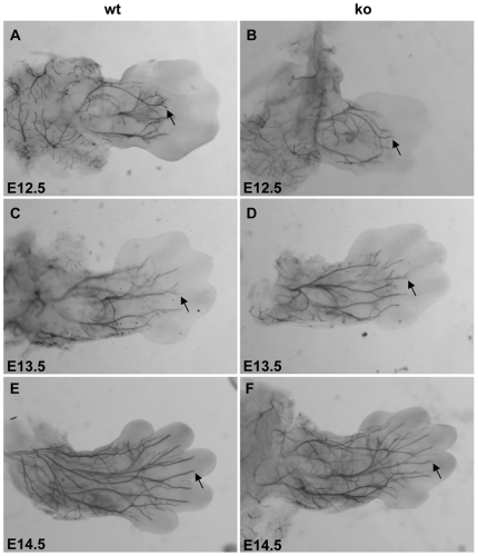
Figure 2. Neurofilament whole-mount immunohistochemistry of the developing limbs in mouse embryos.
Shown is a dorsal view of the forelimbs of wild-type (wt, A, C, E) and palladin-deficient embryos (ko, B, D, F) at ages E12.5 (A, B), E13.5 (C, D), and E14.5 (E, F). Arrows indicate immunoreactive axons projecting into the forelimbs (magnification, 40×).

