Fig. 4.
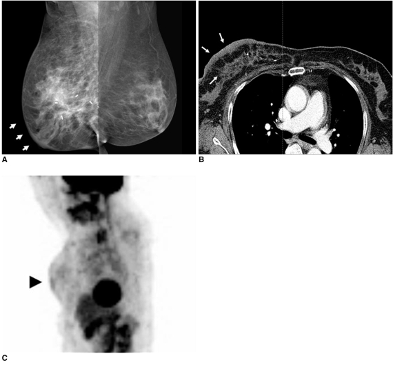
53-year-old female patient who had undergone radiation therapy in right breast six months ago are shown.
A. Mammography of right breast shows diffusely increased density due to skin and trabecular thickening (arrows).
B. Chest CT scan shows diffuse breast edema with skin and trabecular thickening (arrows).
C. Whole body PET image shows diffuse skin thickening and interstitial edema with mild hypermetabolic activity (max SUV = 1.8) (arrowhead) in irradiated right breast.

