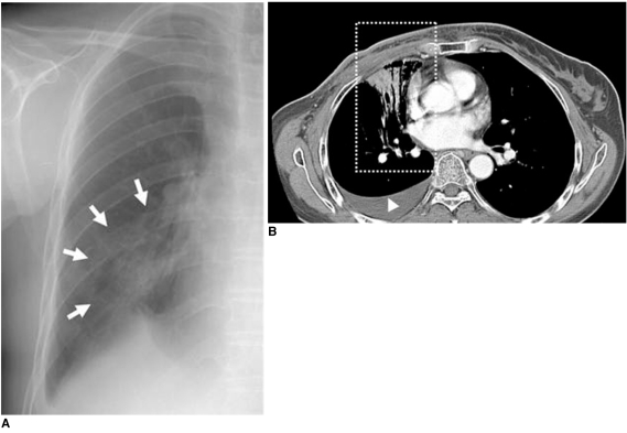
Fig. 8.
52-year-old female patient who had undergone radiation therapy in right breast two weeks ago are shown.
A. Chest radiograph shows peribronchial consolidation in right mid lung field (arrows).
B. Chest CT scan shows air-space consolidation with air-bronchogram localized in medial portion of right lung (box) that was related to previous radiation field for internal mammary lymph nodes, suggesting presence of radiation-induced pneumonia. Associated right pleural effusion is also noted (arrowhead).

