Figure 1.
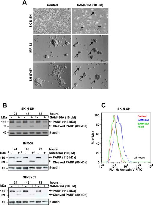
SAM486A induces apoptosis in p53-wild type NB Cells. SK-N-SH, IMR-32, and SH-SY5Y cells were exposed to 10 μM SAM486A or left untreated for 24, 48, and/or 72 hours. A, representative light micrographs demonstrate the effects of SAM486A on the cell morphology (black arrows) after 72 hours. B, whole cell lysates were analyzed by Western blot for PARP cleavage, a marker for late apoptosis, after 24, 48, and 72 hours. C, SK-N-SH cells were analyzed with flow cytometry and annexin V staining, an early apoptosis marker, to confirm the induction of apoptotic cell death after 24 hours. Spd (10μM) reversed SAM486A-induced apoptosis. Similar results (without Spd control) were obtained after 48 and 72 hours (not shown). Data are representative of three independent experiments (n=3). Spd control in C (n=2).

