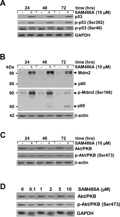
Figure 3.
Analysis of apoptosis-regulating proteins in SAM486A-treated NB cells. Whole cell lysates were obtained from SK-N-SH cells exposed to 10 μM SAM486A for 24, 48, and 72 hours or exposed to increasing concentrations of SAM486A (0, 0.1, 1, 2, 5, and 10 μM) for 72 hours, and analyzed by Western blot. Resolved proteins were transferred to PVDF membranes and sequentially probed for (A) total p53, phospho-p53 (Ser392), and phospho-p53 (Ser46), (B) total Mdm2 and phospho-Mdm2 (Ser 166), (C-D) total Akt/PKB and phospho-Akt/PKB (Ser473). SAM486A treatment increased total p53, phospho-p53 (Ser392), phopho-p53 (Ser46), total Mdm2, phospho-Mdm2 (Ser166) and decreased total Akt/PKB and phospho-Akt/PKB (Ser473). Total Akt/PKB and phospho-Akt/PKB (Ser473) also decreased in a dose-dependent manner. GAPDH or β-actin served as loading markers. Data are representative of three independent experiments (n=3).

