
Table 5.
Corresponding BPh interactions observed in the 3D structures of E. coli and T. thermophilus 16S and 23S rRNAs
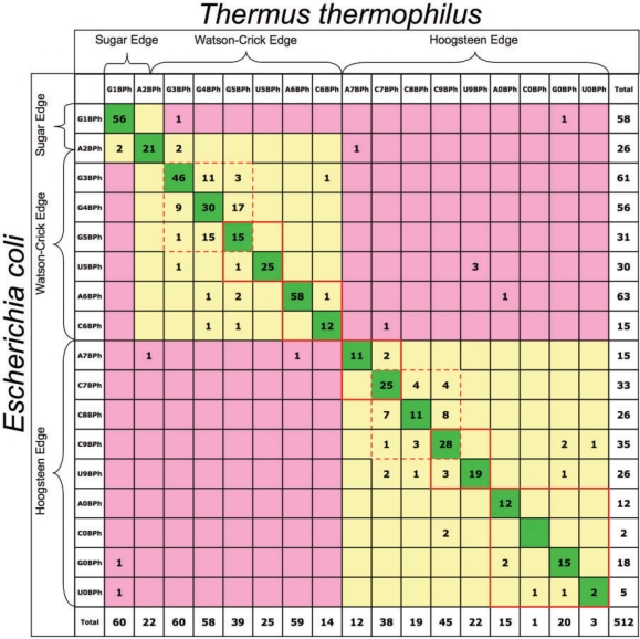 |
Diagonal entries (dark green) correspond to identical BPh interactions (same base donor and BPh category). Yellow shaded cells correspond to differences in base or BPh category that preserve the geometry of the interaction. Pink cells indicate differences that do not preserve the BPh geometry.