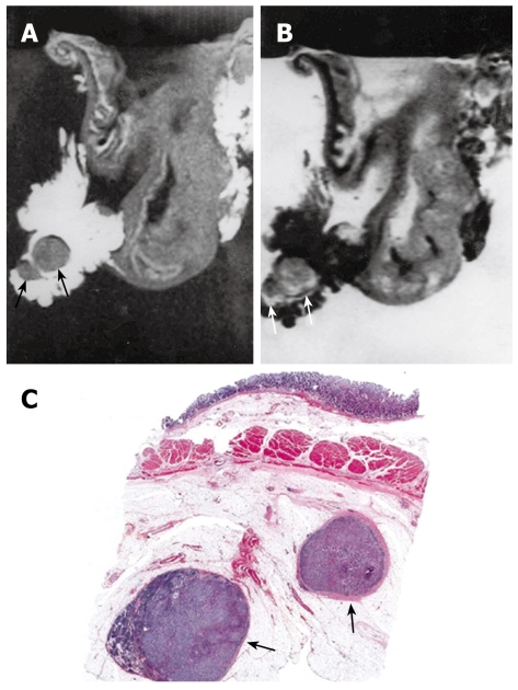
Figure 6.
MRI and histology of N2 gastric adenocarcinoma. A: T1-weighted (500/20) MRI showed two lymph nodes in lesser curvature site of stomach antrum (arrows). Eight lymph nodes are detected in total in perigastric area (not shown); B: T2-weighted (2000/90) MRI showed intermediate SI in the lymph nodes (arrows); C: Light microscopic section showed two lymph nodes in lesser curvature site of gastric antrum (arrows) (HE stain; original magnification, × 1).

