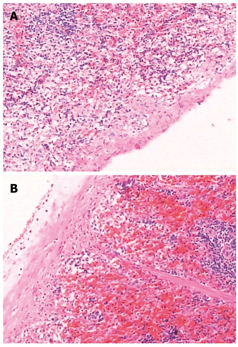
Figure 7.
Pathologic image of the spleen. A: Group-I; B: Group-II, the capsule was thickened, the spleen sinus was enlarged due to congestion, there was significant sinus endothelial cell proliferation, and the spleen trabeculae were widened. The white pulp and germinal center had significant atrophy.

