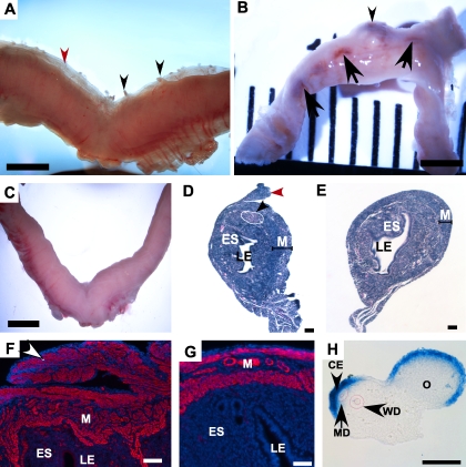
FIG. 1.
Constitutive activation of β-catenin in uterine mesenchyme. A) Gross uterus from Amhr2tm3(cre)Bhr;Ctnnb1tm1Mmt/+ nulliparous mouse showing abnormal growth (black and red arrowheads) on the anti-mesometrial side of the uterus. Nodules were also observed on the surface of the uterus (black arrowheads). B) Gross uterus from multiparous adult Amhr2tm3(cre)Bhr;Ctnnb1tm1Mmt/+ mouse showing tumorous growth (black arrowhead) and multiple hemorrhagic sites (black arrows). C) Normal uterus from a control Amhr2tm3(cre)Bhr/+ mouse. H&E-stained section of 4-wk-old mutant uterus (D) showing polyp-like growth protruding from the myometrium on the anti-mesometrial surface of the uterus (red arrowhead) and ESS-like lesions in the endometrial stroma (black arrowhead) and control uterus (E). ACTA2-immunostained longitudinal section of a nulliparous mutant uterus (F) with positively stained polyps (white arrowhead) and control (G). H) In situ hybridization in the E13.5 urogenital ridge shows that the Amhr2 mRNA is expressed in the coelomic epithelium and subjacent mesenchyme of the Müllerian duct (outlined in red with the Wolffian duct). CE, coelomic epithelium; MD, Müllerian duct; WD, Wolffian duct; O, ovary; LE, luminal epithelium; ES, endometrial stroma; M, myometrium. Bars = 2 mm (A–C), 50 μm (D–H).

